Certification
CarnaLife Holo is certified as a Class IIa diagnostic support medical device by TÜV NORD Polska Sp. z o.o., a notified body authorized by the Ministry of Health.
We also have a NATO Commercial and Government Entity Code (NCAGE)



How does CarnaLife Holo work
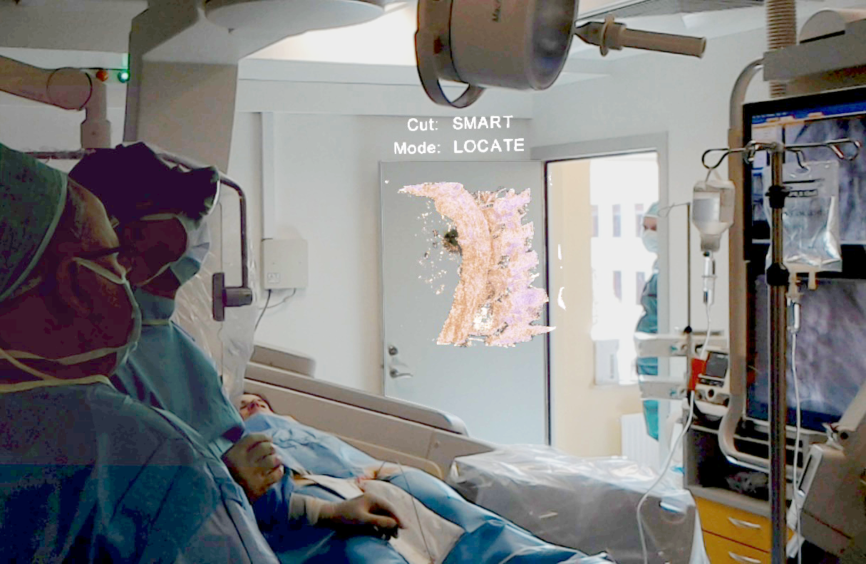
CarnaLife Holo makes it possible to visualize various types of anatomical structures, such as the brain, liver, heart, lungs, bone structures, kidneys, or blood vessels, imaged using various medical imaging techniques recorded in the DICOM standard, such as computed tomography (CT), magnetic resonance imaging (MRI/MR), rotational angiography (3DRA), and three-dimensional echocardiography (ECHO). It is an extremely useful tool for three-dimensional visualization of anatomical structures in the fields of cardiology, interventional cardiology, oncology, orthopedics, general surgery, ENT, pulmonology or interventional radiology, among others. The data can be presented as volumetric, surface and MPR (Multi-Planar Reconstruction) visualization.
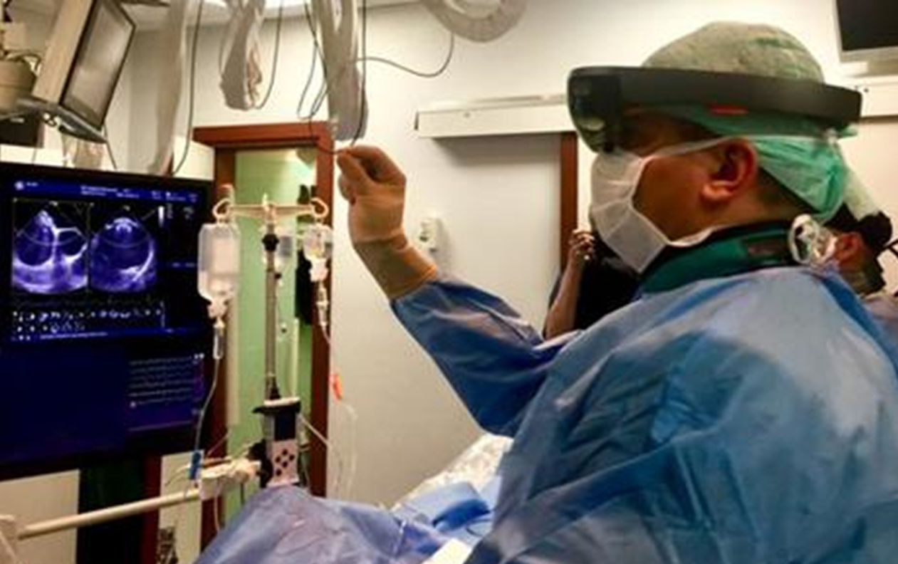
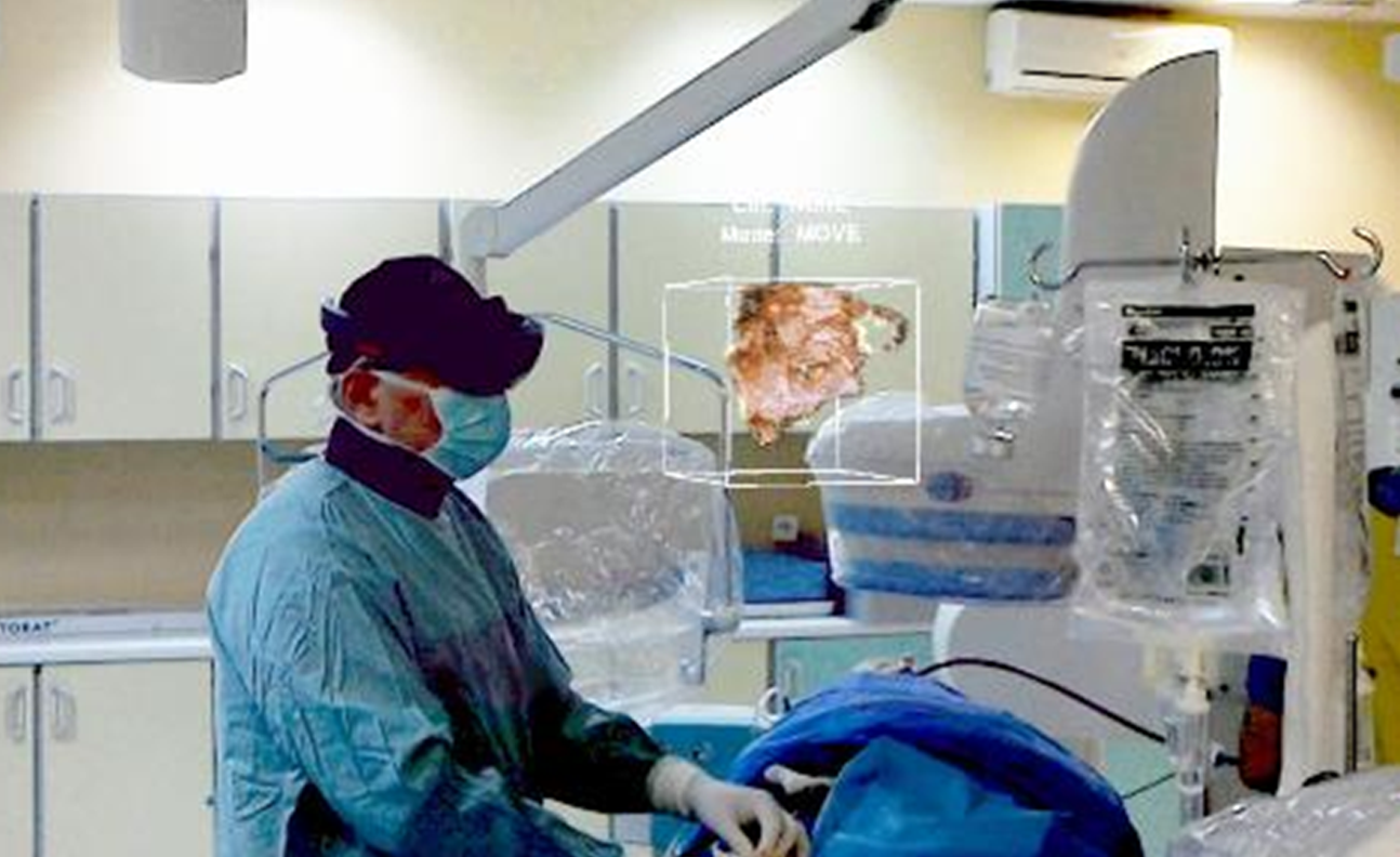
Using our own medical data visualization module based on proprietary algorithms, the system can connect directly with imaging devices in order to display holograms during the examination. An example is working with GE echocardiography devices (Vivid E95), where CarnaLife Holo can provide real-time 3D visualization of data from the ultrasound transducer and display it as interactive holograms. Such an approach ensures that clinical conditions can be independently verified by the operator.
CarnaLife Holo works with hospital PACS (Patient Archiving And Communication System), which makes it possible to visualize the data directly from the hospital IT system, ensuring continuity of the physician’s work.
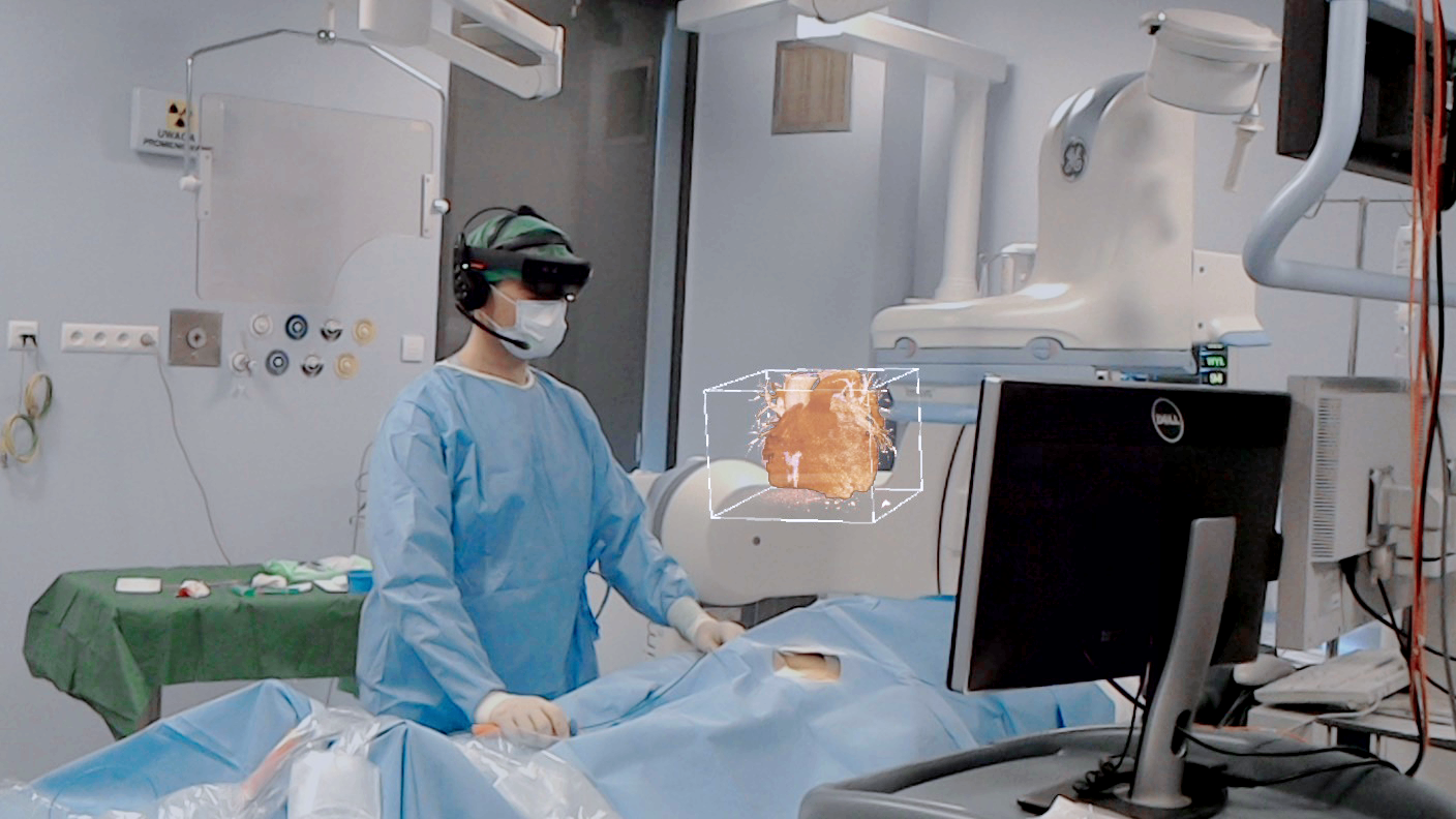
Visualizations of medical images are presented as holograms thanks to Microsoft Hololens 2 mixed reality goggles. The user can interact with the displayed image using gestures and voice commands: rotate it, scale it, move it around, or even go inside the anatomical structures, without compromising sterility or having to work with an additional technician. The system also offers a number of features and tools, including scene customization, filtering, scissors to remove unnecessary elements, built-in presets (custom predefined sets of visualization parameters tailored to specific imaging techniques), and the option to create new effects for specific applications.
The goggles are an additional, interactive screen available during the procedure at any place of the treatment room and at any time.

