A Breakthrough Technology for 3D Visualization of Imaged Diagnostic Data
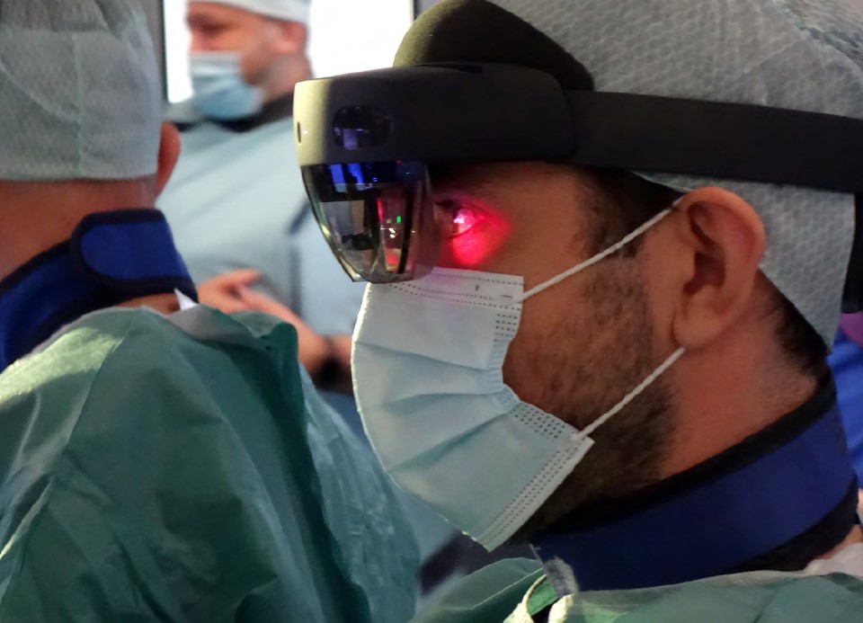
CarnaLife Holo supports the planning and execution of medical procedures. With the help of Microsoft’s HoloLens 2 goggles, the doctor sees in real space a 3D hologram that reflects the structure of the imaged anatomical area. CarnaLife Holo works with hospital PACS (Patient Archiving And Communication System), which makes it possible to show visualizations directly from the hospital IT system, ensuring workflow continuity for the physician.
CarnaLife Holo works well for a variety of teams within a single hospital and is suitable for physicians of any specialty who are already using or would like to implement 3D imaging in their practice. An example taken from the wide range of surgical specialties that can benefit from mixed reality-based technology is vascular surgery of lymphatic and blood vessels.
Surgical Procedure
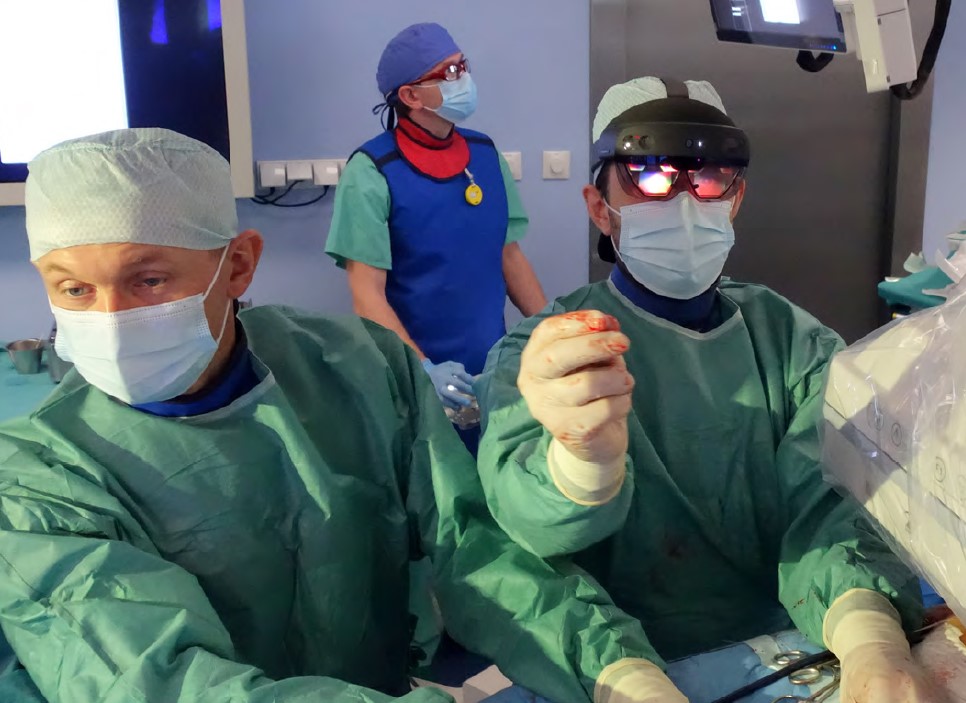
A team of vascular surgeons from the Department of Vascular Surgery and Angiology at the Independent Public University Hospital No. 1 in Lublin, as the first in this part of Poland, successfully performed a novel operation on a patient with an abdominal aortic aneurysm. The procedure took place without unexpected complications. Experienced surgeons were supported by mixed reality at all times, which enabled them to constantly access the patient’s imaged medical data extracted from the CT scan and visualized as a hologram. The technology was useful for both the surgeons and the patient, indirectly reducing the duration of the surgery, which took about an hour. The patient tolerated the procedure very well and was under epidural anesthesia the entire time.
Parameterizing the Aneurysm Dimensions
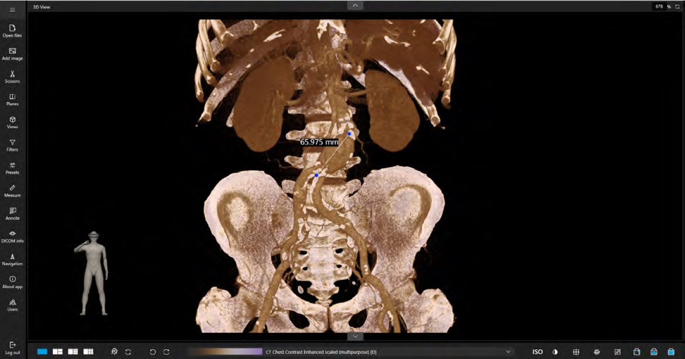
An important element of the performed procedure was the ability to parameterize the dimensions of the aneurysm located in the patient’s abdominal aorta. CarnaLife Holo has a functionality that measures anatomical structures. This feature is already medically certified for diagnostic purposes and enables:
• Unique, instantaneous conversion of measurements from 2D to 3D and holographic space.
• “Cut Smart” feature for making any plane in the hologram visible using head movements.
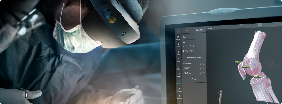
• Representation of measurements in holographic space. • Annotation: marking points of interest in the medical data that can be easily found in the holographic space. This approach makes it possible to highlight the next steps of the procedure and annotate in real time.
• Customization options: display measurements according to the surgeon’s needs with different sizes, colors and modes.
Abdominal Aortic Aneurysms
The number of abdominal aortic aneurysms (AAA) diagnosed and operated on is increasing every year. AAA’s are most common in people over 65 years of age.1 It is considered that they occur in 2-4% of people under 65 and in about 9% of people over 75 years of age.2 The presence of abdominal aortic aneurysms in the elderly is directly related to the half-life of elastin, which is 70 years. The incidence of abdominal aortic aneurysms in men is 3 to even 9 times higher than in women.3 The main risk factors for AAA are age over 65, male sex, smoking, genetic predispositions, hypertension, and emphysema.4
1. Steckmeier B: Epidemiology of aortic disease: aneurysm, dissection, occlusion. Radiologe 2001; 41(8): 624-632. 2. Ligush J Jr, Pearce JD, Edwards MS et al.: Analysis of medical risk factors and outcomes in patients undergoing open versus endovascular abdominal aortic aneurysm repair. J Vasc Surg 2002; 36(3): 492-499. 3. Clouse WD 2. Clouse WD, Hallett JW Jr, Schaff HV: Acute aortic dissection: population-based incidence compared with degenerative aortic aneurysm rupture. Mayo Clin Proc 2004; 79(2): 176-180. 4. Prisant LM, Mondy JS 3rd: Abdominal aortic aneurysm. J Clin Hypertens (Greenwich) 2004; 6(2): 85-89. 3. Collin J, Araujo L, Walton J et al.: Oxford screening programme for abdominal aortic aneurysm in men aged 65 to 74 years. Lancet 1988; 2(8611): 613-615. 4. https://bit.ly/3xRLkHG
Summary
To visualize a hologram of the operated abdominal aorta, IPUH1 surgeons used CarnaLife Holo and Microsoft’s HoloLens 2 goggles. In real space, they saw a three-dimensional hologram that accurately depicted the anatomical fragment being operated on. The ability to interact with the displayed hologram, including rotation, zooming in or out while maintaining sterility, was described as a breakthrough in current practice by the Lublin surgeons. They said that not only is it easier for the surgeon to operate, but that patient’s safety is also increased. The hologram helps to avoid mistakes in complicated procedures
In this patient’s case, CarnaLife Holo successfully helped to support a life-saving procedure: implanting a stent graft into the patient’s aorta without the use of a scalpel. Early diagnosis was also crucial to avoid uncontrolled aortic aneurysm rupture. The 3D medical data visualization for surgical interventions is already being used successfully in cardiology, interventional cardiology, orthopedics, otorhinolaryngology, as well as oncologic surgery. Treatment at Lublin’s Independent Public University Hospital No. 1 in Lublin has added vascular surgery to the portfolio of specializations using CarnaLife Holo technology